
Colorectal Cancer Guide

Blood tumour markers for monitoring colorectal cancer

CEA
Carcinoembryonic antigen (CEA) is a protein produced by tumour cells that can be measured in the blood. In colorectal cancer, CEA levels are often higher than normal. Although CEA is not used to diagnose colorectal cancer, it is an important biomarker for monitoring the disease over time, helping assess how well treatment is working and whether the cancer may have returned1.
After treatment, CEA levels are often measured regularly during follow-up. A decrease in CEA levels usually suggests that treatment is working, while stable or rising levels may indicate that cancer is still present or has returned. However, CEA has important limitations. CEA levels can be affected by non-cancerous conditions, and a normal or raised CEA level alone cannot confirm whether cancer has or has not returned. For this reason, CEA results must always be interpreted alongside scans and clinical evaluation1.
Biomarkers that help guide treatment
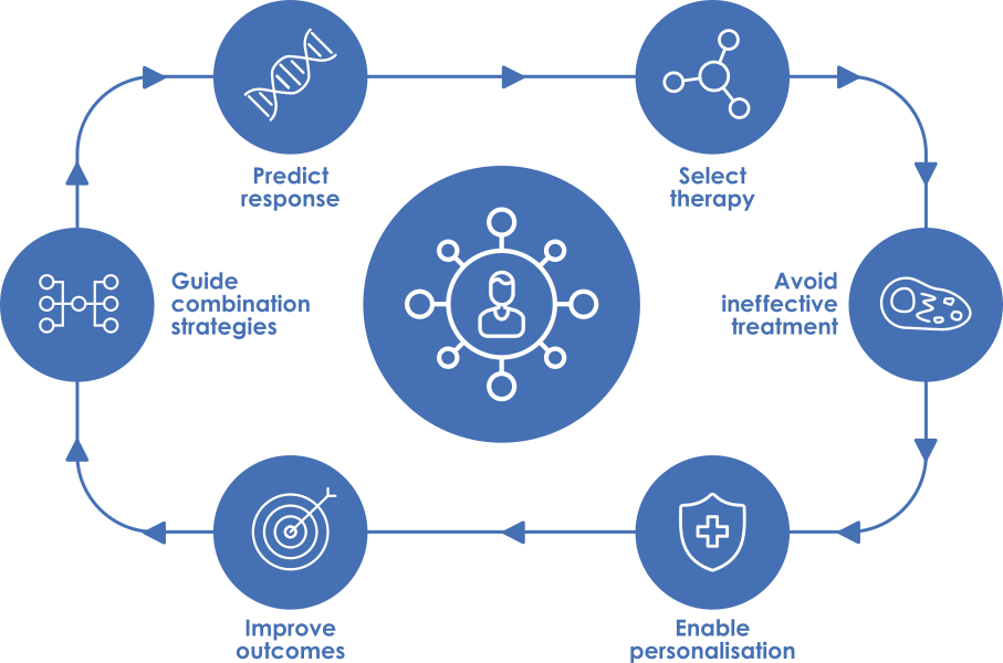
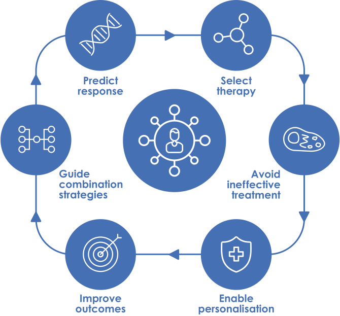
All patients with colorectal cancer, regardless of stage, should undergo testing for microsatellite instability (MSI) or mismatch repair deficiency (dMMR), as this provides key information for both prognosis and treatment selection. In metastatic disease (stage IV), additional testing is recommended for KRAS, NRAS, BRAF and HER2 to optimise therapy selection2. According to guidelines, molecular testing should be performed in all patients at the time of metastatic diagnosis, either on the primary tumour or metastatic tissue, and results should ideally be available within 14 days (but the number of days heavily relies on the laboratory’s workload and many other factors)2.
MSI reflects the inability of tumour cells to repair DNA damage. When the mismatch repair (MMR) system is functioning normally, tumours are classified as microsatellite stable (MSS). When this system is defective due to alterations in genes such as MLH1, MSH2, MSH6, or PMS2, mutations accumulate and the tumour becomes MSI-high (MSI-H) or dMMR1,3. These tumours account for approximately 15% of colorectal cancers, although they are less common in metastatic disease, and rare in rectal cancer1,3.
MSI-H or dMMR tumours are clinically important because they tend to respond particularly well to immunotherapy, especially immune checkpoint inhibitors such as pembrolizumab or nivolumab2,3. While most cases are caused by somatic tumour-specific changes, a minority are associated with germline mutations linked to Lynch syndrome. In these cases, identification of MSI-H status has implications beyond the individual patient, as it may indicate an inherited cancer predisposition, making genetic counselling important for both patients and their families2,4,5.
RAS genes, including KRAS and NRAS, play a central role in regulating cell growth. KRAS and NRAS mutations are common in colorectal cancer, occurring in approximately 40% and 4% of patients respectively, and have major therapeutic implications1,6,7. Tumours with RAS mutations do not respond to anti-EGFR therapies such as cetuximab or panitumumab5. In contrast, patients without RAS mutations (wild-type) tumours may benefit from these treatments3,8.
The Raf murine sarcoma viral oncogene homolog B (BRAF) protein is produced when cells need to grow. An abnormal production of BRAF can cause the cells to grow continuously, leading to cancer development. BRAF mutations, most notably the V600E mutation, occur in 8-12% of metastatic cases and are associated with more aggressive disease, poorer prognosis, and reduced response to standard therapies, particularly when anti-EGFR agents are used alone3,5. However, targeted combinations such as BRAF inhibitors with other agents (e.g., encorafenib and cetuximab) have shown improved outcomes2. Importantly, patients with both BRAF mutations and MSI-H status may also benefit from immunotherapy, underlining the importance of comprehensive molecular profiling5.
Human epidermal growth factor receptor 2 (HER2) is a protein that can promote cancer cell growth. HER2 is overexpressed or amplified in a small subset of colorectal cancers, approximately 3% of colon cancers and 8% of rectal cancers, and is more common in patients without RAS mutations (wild-type)3,8. Although it is associated with resistance and poor response to anti-EGFR therapies (such as cetuximab and panitumumab), it also identifies patients who may benefit from HER2-targeted treatments2. HER2 mutations can also be acquired over time as a resistance mechanism to therapies such as EGFR inhibitors, so repeating HER2 testing at disease progression or recurrence can be useful9.
Less common but highly relevant biomarkers include Neurotrophic receptor kinase (NTRK) gene fusions and mutations in DNA polymerase epsilon (POLE) or DNA polymerase delta 1 (POLD1). Although rare (<1%), these alterations are clinically important because they can predict strong responses to targeted therapies or immunotherapy. NTRK gene fusions can be treated with TRK inhibitors such as larotrectinib or entrectinib2. POLE and POLD1 mutations are associated with tumours that have a very high number of genetic changes and may respond particularly well to immunotherapy drugs called immune checkpoint inhibitors2.
In addition, phosphatidylinositol-4,5-bisphosphate 3-kinase catalytic subunit alpha (PIK3CA) mutations occur in around 10-15% of cases and may be associated with resistance to anti-EGFR therapy1,3,5.
Beyond molecular features, the physical location of the tumour within the colon, also carries important clinical meaning, influencing both prognostic and therapeutic implications.
- Right-sided colorectal tumours arise in the first part of the colon and are more often associated with MSI-H/dMMR status, as well as mutations in genes such as KRAS and BRAF, and less commonly NTRK rearrangements2. These tumours are often diagnosed at more advanced stages and generally have a poorer prognosis compared with left-sided cancers2. Because of their molecular profile, right-sided tumours are less responsive to anti-EGFR therapies2. As a result, treatment in metastatic disease often involves chemotherapy combined with antiangiogenic agents, unless MSI-H/dMMR status suggests that immunotherapy may be a better option3.
- Left-sided colorectal tumours tend to present earlier, have a better prognosis, and are more likely to respond to anti-EGFR therapies, particularly in patients without RAS mutations (wild-type)2,3.
Overall, tumour location is an important factor in guiding first-line treatment decisions, with right-sided disease favouring chemotherapy plus antiangiogenic agents and left-sided disease favouring chemotherapy plus anti-EGFR therapy2,3. This distinction, together with molecular profiling, helps guide more personalised treatment strategies.
Biomarkers that improve treatment safety
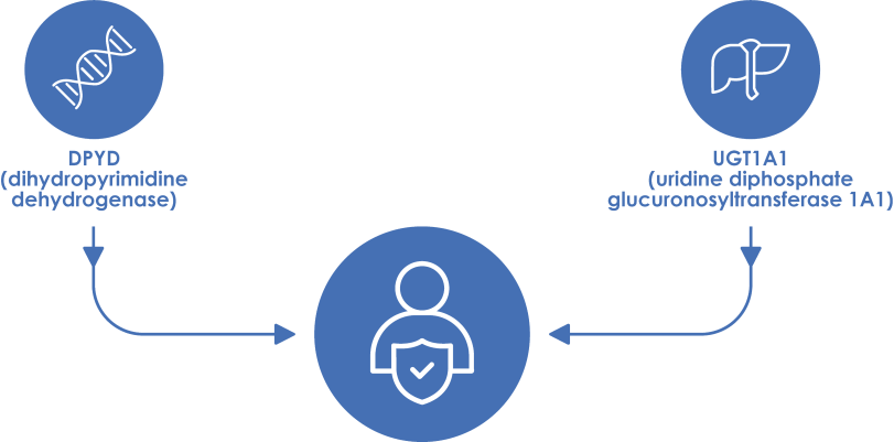
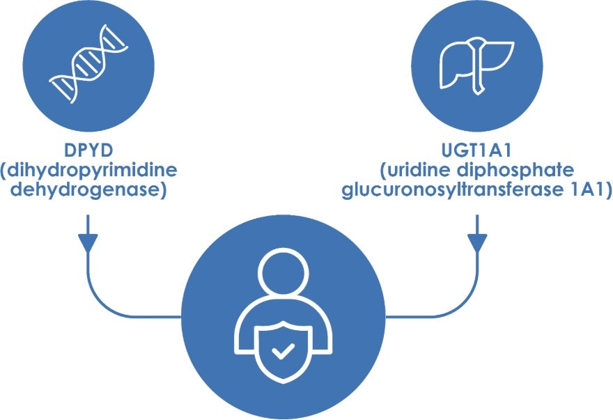
In addition to predicting treatment response, certain biomarkers should be tested before chemotherapy since they are important for ensuring treatment safety. These tests can identify patients who may not break down certain drugs in the usual way, which could increase the risk of serious side effects.
One of the most relevant is the DPYD gene, which helps the body break down chemotherapy drugs such as 5-fluorouracil (5-FU) and related drugs (capecitabine, FOLFOX, FOLFIRI or FOLFIRINOX). Some people have changes in this gene that reduce how well the drug is processed. When this happens, the drug can build up in the body and lead to more severe side effects than expected, sometimes affecting the digestive system, blood cells, or overall energy levels3. For this reason, DPYD testing is recommended before starting treatment, allowing dose adjustment or alternative therapies when necessary2.
Similarly, the UGT1A1 gene is involved in how the body processes irinotecan, a commonly used chemotherapy drug. Certain changes in this gene can slow down drug breakdown, meaning the drug stays in the body longer. This can increase the chance of stronger side effects, so testing can help doctors decide whether dose adjustments are needed to improve safety11.
1. Zamanian, M. Y. et al. Biomarkers for colorectal cancer detection: An insight into colorectal cancer and FDA-approved biomarkers. BioImpacts 15, 31211 (2025).
2. Cremolini, C. et al. Metastatic colorectal cancer: ESMO Clinical Practice Guideline for diagnosis, treatment and follow-up. Annals of Oncology https://doi.org/10.1016/j.annonc.2026.03.005 (2026) doi:10.1016/j.annonc.2026.03.005.
3. Patel, J. N., Fong, M. K. & Jagosky, M. Colorectal Cancer Biomarkers in the Era of Personalized Medicine. J. Pers. Med. 9, 3 (2019).
4. Lișcu, H.-D. et al. Biomarkers in Colorectal Cancer: Actual and Future Perspectives. Int. J. Mol. Sci. 25, 11535 (2024).
5. Sepulveda, A. R. et al. Molecular Biomarkers for the Evaluation of Colorectal Cancer. The Journal of Molecular Diagnostics 19, 187–225 (2017).
6. Zhang, Y., Wang, Y., Zhang, B., Li, P. & Zhao, Y. Methods and biomarkers for early detection, prediction, and diagnosis of colorectal cancer. Biomedicine & Pharmacotherapy 163, 114786 (2023).
7. Levin-Sparenberg, E. et al. A Systematic Literature Review and Meta-Analysis Describing the Prevalence of KRAS, NRAS, and BRAF Gene Mutations in Metastatic Colorectal Cancer. Gastroenterology Res. 13, 184–198 (2020).
8. Hakami, Z. H. Biomarker discovery and validation for gastrointestinal tumors: A comprehensive review of colorectal, gastric, and liver cancers. Pathol. Res. Pract. 255, 155216 (2024).
9. Harrold, E. et al. Molecular and Clinical Determinants of Acquired Resistance and Treatment Duration for Targeted Therapies in Colorectal Cancer. Clinical Cancer Research 30, 2672–2683 (2024).
10. Abbas, N., Mourad, M., Smaily, H., Al Mahmasani, L. & Shamseddine, A. EGFR Signaling in Colorectal Cancer: Novel Therapeutic Strategies, Predictive Biomarkers, and Counteracting Treatment Resistance. Int. J. Mol. Sci. 27, 3265 (2026).
11. Karas, S. & Innocenti, F. All You Need to Know About UGT1A1 Genetic Testing for Patients Treated With Irinotecan: A Practitioner-Friendly Guide. JCO Oncol. Pract. 18, 270–277 (2022).